Question 15#
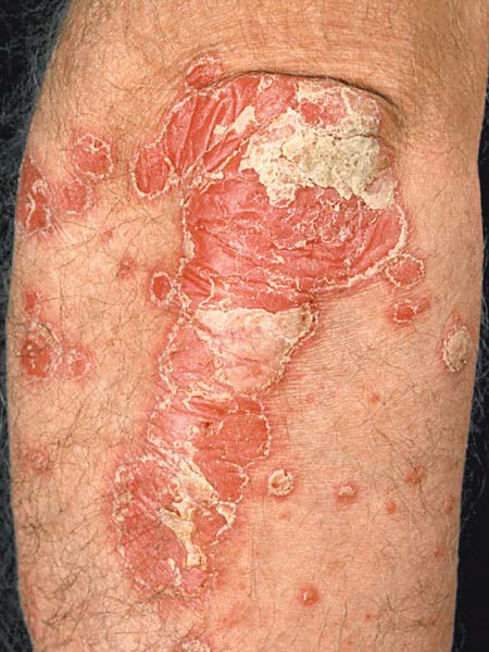
A 53-year-old woman presents with pain in the fingers bilaterally. Examination reveals inflammation of the synovium of multiple DIP and PIP joints. Larger joints are spared. Skin examination reveals the following lesion (pic).
What is the most likely diagnosis?
A. Hemochromatosis
B. Rheumatoid arthritis
C. Osteoarthritis
D. Systemic lupus erythematosus
E. Psoriatic arthritis
Correct Answer is E
Comment:
The patient has psoriatic arthritis. Psoriatic arthritis is an immune-mediated arthritis affecting up to 30% of patients with psoriasis. Although psoriasis usually precedes the development of arthritis, occasionally the joint symptoms come first. Classic manifestations include involvement of the DIP joints, presence of dactylitis (“sausage digit”), and of course the presence of psoriasis of the skin. Joint involvement patterns are variable, however, and can mimic rheumatoid arthritis (as in this case) or involve primarily the axial skeleton. Nail changes, including pitting, horizontal ridging, onycholysis, dystrophic hyperkeratosis, and yellowish discoloration are common. The diagnosis is primarily clinical, although radiographic evidence of “pencil in cup” deformity of the DIP joint may lend additional weight to the diagnosis. Immunosuppression is required to prevent deformation of the joints, with anti-TNF alpha agents being the current drug of choice. Although hemochromatosis can cause joint involvement, it typically mimics the noninflammatory presentation of osteoarthritis, often with involvement of the second and third MCP joints (an unusual pattern in primary osteoarthritis). The cardinal skin manifestation of hemochromatosis is diffuse hyper-pigmentation, leading to the colloquial description of “bronze diabetes.” Rheumatoid arthritis could cause the joint manifestations described here, but would not result in the portrayed skin lesion. RA classically affects the metacarpophalangeal (MCP) and spares the DIP joints. Osteoarthritis will not present with polyarticular synovitis or a psoriatic plaque. SLE can mimic many diseases, but discoid lupus lesions typically have an atrophic center with erythematous scaly edge, as opposed to the uniformly thick scale seen in psoriasis.