Question 2#
Chronic venous insufficiency ( CVI) is characterized by all of the following EXCEPT:
A. Incompetence of venous valves, venous obstructionB. Preserved microcirculatory and cutaneous lymphatic anatomy
C. Eczema and dermatitis
D. Lipodermatosclerosis
Correct Answer is B
Comment:
Chronic venous insufficiency ( CVI) may lead to characteristic changes in the skin and subcutaneous tissues in the affected limb. CVI results from incompetence of venous valves, venous obstruction, or both. Most CVI involves venous reflux, and severe CVI often reflects a combination of reflux and venous obstruction. It is important to remember that although CVI originates with abnormalities of the veins, the target organ of CVI is the skin, and the underlying physiologic and biochemical mechanisms leading to the cutaneous abnormalities associated with CVI are poorly understood. A typical leg affected by CVI will be edematous, with edema increasing over the course of the day. The leg may also be indurated and pigmented with eczema and dermatitis. These changes are associated with excessive proteinaceous capillary exudate and deposition of a pericapillary fibrin cuff that may limit nutritional exchange. In addition, an increase in white blood cell (WBC) trapping within the skin microcirculation in CVI patients may lead to microvascular congestion and thrombosis. Subsequently, WBCs may migrate into the interstitium and release necrotizing lysosomal enzymes, potentially leading to tissue destruction and eventual ulceration. Fibrosis can eventually develop from impaired nutrition, chronic inflammation, and fat necrosis (lipodermatosclerosis). Hemosiderin deposition due to the extravasation of red cells and subsequent lysis in the skin contributes to the characteristic pigmentation of chronic venous disease (Fig. below). Ulceration can develop with longstanding venous hypertension and is associated with alterations in microcirculatory and cutaneous lymphatic anatomy and function. The most common location of venous ulceration is approximately 3 cm proximal to the medial malleolus (Fig. below).
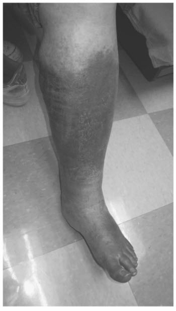
Characteristic hyperpigmentation of chronic venous insufficiency.
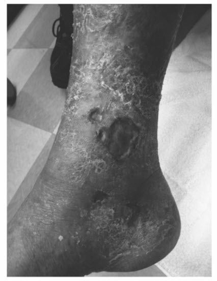
Venous ulceration located proximal to the medial malleolus.