Question 9#
The most common level of cervical radiculopathy from cervical disc herniation is:
A. C4-C5B. C5-C6
C. C6-C7
D. C7-Tl
Correct Answer is C
Comment:
The cervical nerve roots exit the central canal above the pedicle of the same-numbered vertebra and at the level of the higher adjacent intervertebral disc. For example, the C6 nerve root passes above the C6 pedicle at the level of the C5-C6 discs. The cervical nerve roots may be compressed acutely by disc herniation, or chronically by hypertrophic degenerative changes of the discs, facets, and ligaments. Table below summarizes the effects of various disc herniations. Most patients with acute disc herniations. Most patients with acute disc herniations will improve without surgery, nonsteroidal antiinflammatory drugs (NSAIDs), or cervical traction may help alleviate symptoms. Patients whose symptoms do not resolve or who have significant weakness should undergo decompressive surgery. The two main options for nerve root decompression are anterior cervical discectomy and fusion (ACDF) and posterior cervical foraminotomy (keyhole foraminotomy). ACDF allows more direct access to and removal of the pathology (anterior to the nerve root). However, the procedure requires fusion because discectomy causes a collapse of the interbody space and instability will likely occur. Fig. below demonstrates a C6-C7 ACDF with the typical interposed graft and plating system. Keyhole foraminotomy allows for decompression without requiring fusion, but it is less effective for removing centrally located canal pathology.
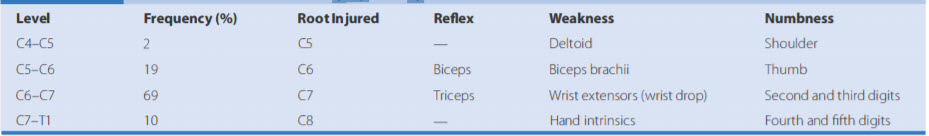
Cervical disc herniations and symptoms by level table:
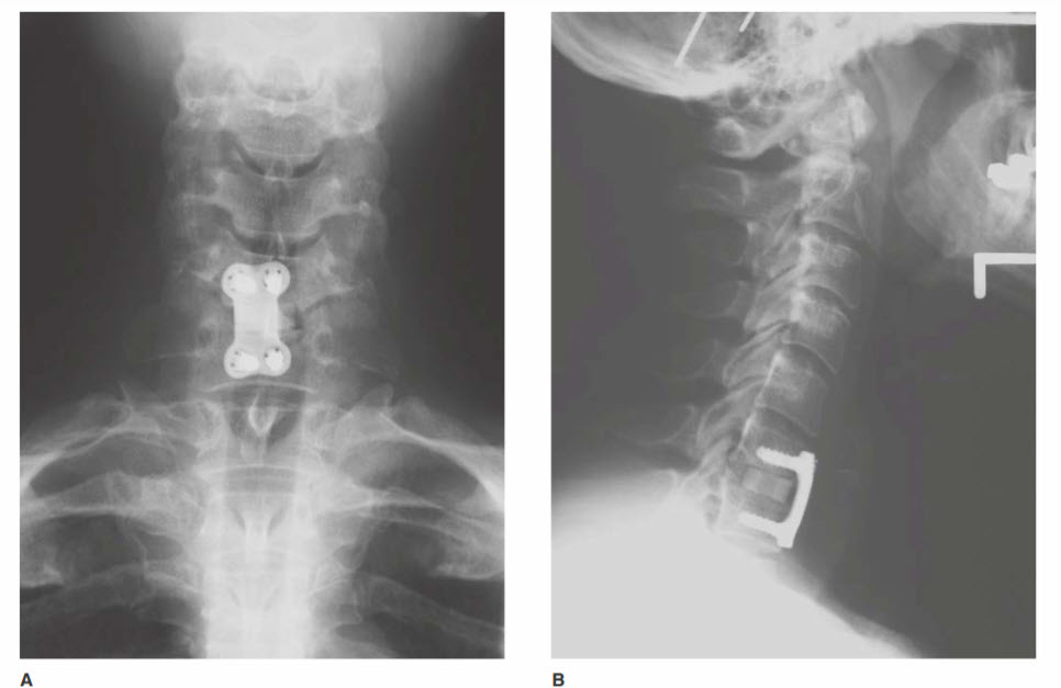
A. Anteroposterior cervical spine X-ray showing the position of an anterior cervical plate used for stabilization after C6-C7 discectomy. Patient presented with right triceps weakness and dysesthesias in the right fifth digit. Magnetic resonance imaging (MRI) revealed a right paracentral C6-C7 herniated disc compressing the exiting C7 nerve root. B. Lateral cervical spine X-ray of the same patient clearly demonstrates the position of the plate and screws. The allograft bone spacer placed in the drilled-out disc space is also apparent.