Question 11#
Which of the following about enchondromas is true?
A. Have never been reported in the trapezoidB. Discovery is often prompted by patients presenting with hand pain
C. Are the most common malignant primary bone tumors
D. The most common location is the middle phalanges
Correct Answer is A
Comment:
This is the most common primary benign bone tumor of the hand and wrist and is of cartilage origin. Up to 90% of all bone tumors in the hand and wrist are enchondromas, with 35 to 54% of all enchondromas occurring in the hand and wrist. They are often found incidentally on X-rays taken for other reasons (eg, hand trauma). They are usually solitary and favor the diaphysis of small tubular bones and are most common in the second and third decades of life. The most common location is in the proximal phalanges, followed by the metacarpals and then middle phalanges. Enchondroma has never been reported in the trapezoid. Presentation is usually asymptomatic, but pain may occur if there is a pathologic fracture or impending fracture. The etiology is believed to be from a fragment of cartilage from the central physis. Histology shows well-differentiated hyaline cartilage with lamellar bone and calcification. Two variants of enchondroma include Ollier disease (multiple enchondromatosis) and Maffucci syndrome (multiple enchondromatosis associated with multiple soft tissue hemangiomas). Malignant transformation is very rare in the solitary form, but there is a 25% incidence by age 40 in Ollier patients and a 100% life-time incidence in Maffucci patients. When malignant transformation does occur, it is almost uniformly a chondrosarcoma with pain and rapid growth. Diagnosis is usually made based on history, physical examination, and X-rays. There is a well-defined, multilobulated central lucency in the metaphysis or diaphysis that can expand causing cortical thinning or sometimes, thickening (Fig. below). Further imaging is seldom needed, but a CT would be the study of choice.
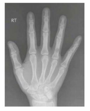
Enchondroma. X-ray of the phalanx demonstrates a welldefined central lucency. Surrounding cortex may thin or thicken. Thinning of the cortex contributes to risk of pathologic fracture.