Question 34#
A laboring patient at 40 weeks’ gestation presents with spontaneous rupture of membranes. Bedside ultrasonography shows no measurable pockets of amniotic fluid. With each contraction, the fetal heart rate tracing shows evidence of umbilical cord compression.
Match the description with the appropriate fetal heart rate tracing. If none of the tracings apply, answer e. (none).
e. None
A. (a)B. (b)
C. (c)
D. (d)
E. (e)
Correct Answer is D
Comment:
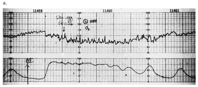
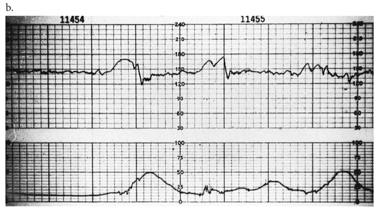
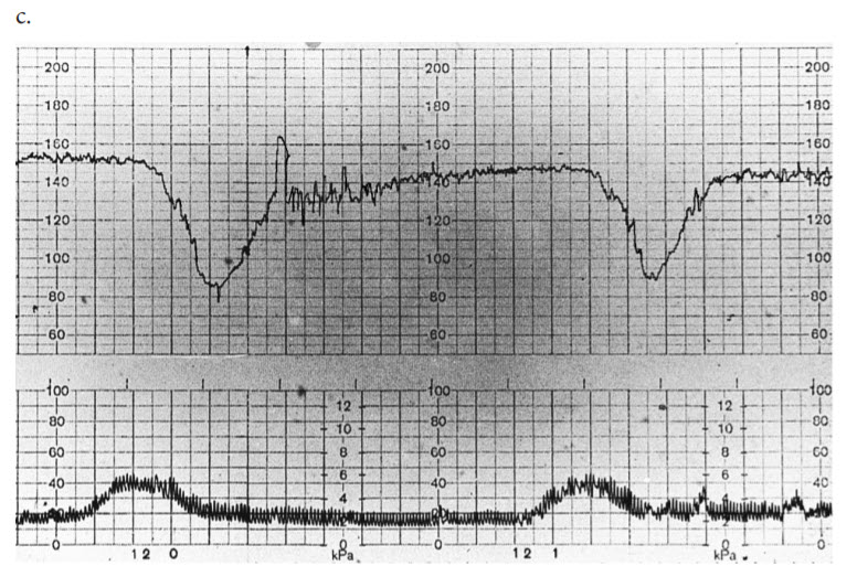
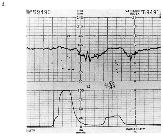
Fetal heart rate tracings are obtained in most pregnancies in the United States through the use of electronic fetal monitoring equipment. Accurate interpretation of these tracings with resultant action to expedite delivery in fetuses threatened by hypoxia has improved neonatal outcome. Electronic fetal monitoring has had very little effect on the overall incidence of cerebral palsy, which seems most often to have its etiology remote from the time of labor. Tracing (a) shows a classic hyperstimulation pattern, with a tonic contraction lasting several minutes with distinctly raised intrauterine pressure and a consequent fall in fetal heart rate. Despite the increased uterine pressure, there remains good beat-to-beat variability, which suggests that the fetus is withstanding the stress. Tracing (b) shows fetal heart rate accelerations occurring spontaneously both before and after contractions, with good beat-to-beat variability, and is representative of a healthy fetus. Tracing (c) shows late decelerations following two consecutive contractions. The baseline variability is significantly reduced. This pattern is caused by uteroplacental insufficiency. Tracing (d) shows variable decelerations in which the classic V-shaped picture of a variable deceleration is maintained. Such decelerations are a normal, reflex response to umbilical cord compression.