Question 3#
A 69-year-old male has undergone a radical cysto-prostatectomy for muscleinvasive bladder cancer with no histological evidence of peri-vesical invasion. Staging investigations included a CT of the chest and abdomen, which showed no metastasis. However, histology of the lymphadenectomy specimen showed that two lymph nodes in the external iliac group were positive for metastasis. What would be the correct TNM (2017) pathological staging?
A. pT2 pN1 M0B. pT3 pN1 M0
C. pT2 pN2 M0
D. pT2 pN3 M0
Correct Answer is C
Comment:
Answer C
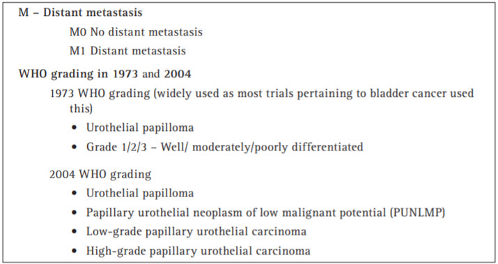
The Tumour, Node, Metastasis (TNM) Classification of Malignant Tumours is the method most widely used to classify the extent of cancer spread. An eighth edition was published in 2017.
2017 TNM Classification of Urinary Bladder Cancer:
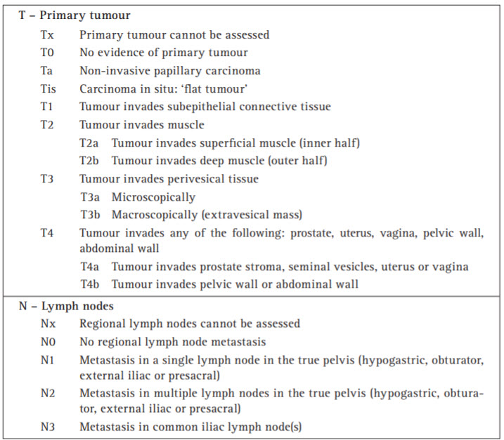
Both CT and MR imaging may be used for assessment of local invasion, but they are unable to detect microscopic invasion of perivesical fat (T3a). The aim of CT and MR imaging is therefore to detect T3b disease or higher. The assessment of nodal status based simply on size is limited by the inability of both CT and MR imaging to identify metastases in normal sized or minimally enlarged nodes. Pelvic nodes greater than 8 mm and abdominal nodes greater than 10 mm in maximum short axis diameter should be regarded as enlarged on CT and MR imaging.
Sensitivities for detection of lymph node metastases are low, ranging from 48% to 87%. Specificities are also low as nodal enlargement may be due to benign pathology. The pN (pathological Node) category is closely related to the number of lymph nodes studied by the pathologist. For this reason, some authors have observed that more than nine lymph nodes have to be investigated to reflect pN0 appropriately after cystectomy.
Further Reading:
- EAU Guidelines on Bladder Cancer Muscle-invasive and Metastatic. Chairman A. Stenzl
- Oyen RH, Van Poppel HP, Ameye FE, et al. Lymph node staging of localized prostatic carcinoma with cT and cT-guided fine-needle aspiration biopsy: Prospective study of 285 patients. Radiology 1994; 190(2): 315–322.
- Barentsz JO, Engelbrecht MR, Witjes JA, et al. MR imaging of the male pelvis. Eur Radiol 1999; 9(9): 1722–1736.