Question 34#
The ECG in the figure below:
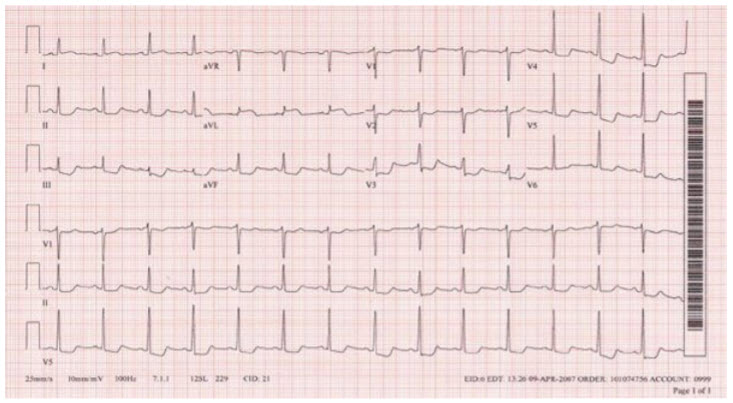
is suggestive of:
A. Acute Anterolateral infarctB. Acute high lateral infarct
C. Acute Anteroseptal infarct
D. Acute Anterior infarct
Correct Answer is B
Comment:
Acute high lateral infarct. There is a sinus rhythm with ST-segment elevation in lead aVL and reciprocal changes in the inferior and lateral leads. This is an acute high lateral infarct.